Simply so, when can you see an embryo on ultrasound?
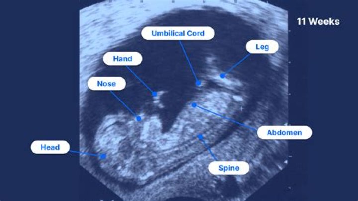
By 8 weeks gestation, your baby can usually be seen with transabdominal ultrasound. By 10-11 weeks gestation, the embryo is clearly recognisable as a baby with a body, head, arms and legs, as well as many other identifiable features. Your baby may be moving around the pregnancy sac.
Subsequently, question is, how many times do you get an ultrasound during pregnancy? Most healthy women receive two ultrasound scans during pregnancy. "The first is, ideally, in the first trimester to confirm the due date, and the second is at 18-22 weeks to confirm normal anatomy and the sex of the baby," explains Mendiola.
Hereof, can you be pregnant and not see the baby on an ultrasound?
Having a gestational sac does not say much about the health of your pregnancy, nor does it say whether an embryo is present or not. If a gestational sac is not seen on an early pregnancy transvaginal ultrasound by around 5 weeks gestational age, there are several things that could be occurring.
What can you see on a 5 week ultrasound?
This week, a sonographer can see your baby via ultrasound as a tiny white image tucked within the gestational sac. Your embryo now looks less like a ball and more like a curled tube. One end of it will eventually become your baby's head; the other, your baby's bottom.
Related Question Answers
Is it common not to see an embryo at 6 weeks?
An embryo is usually seen within the gestational sac by 6 weeks gestation. One of the more common types of miscarriages, known as an anembryonic pregnancy,4? empty sac, or blighted ovum, happens when a gestational sac does not contain an embryo. In other words, an embryo failed to develop.Is it normal to not see an embryo at 5 weeks?
Since the general definition of a blighted ovum is that there's no fetal pole visible on a 7-week ultrasound, this kind of miscarriage can be misdiagnosed if you're not as far along as you think — after all, if you're only 5 weeks pregnant instead of 6 or 7, it's normal to not quite be able to see your teeny-tiny babyWhat kind of ultrasound is done at 7 weeks?
A transvaginal ultrasound finds the heartbeat fairly early, usually between 6 and 7 weeks of gestation.What does a baby look like at 6 weeks?
At six weeks pregnant, your baby's body is taking on a C shape, and small buds that will become his arms and legs are visible. His tiny facial features, including his eyes, nose, ears, chin and cheeks, are also beginning to form. Your baby is about the size of a sweet pea now, and he'll double in size again next week.What can I expect to see at a 7 week ultrasound?
A strong fetal heartbeat can be clearly seen at 7 weeks. The range can be from 100 to 180 beats per minute (bpm) . Any earlier than 7 weeks, you may not see the embryo or fetal heart beating due to the embryo being so small. A gestational sac and yolk sac may only be visible.How early can a pregnancy be detected?
It is recommended that you wait until you have missed a period to take a home test. A missed period is often one of the first signs of pregnancy. If you cannot wait that long to find out and you know the day you may have conceived, then the earliest you can take a test would be 14 days from possible conception.Is it normal not to hear a heartbeat at 6 weeks?
If no heartbeat is detected, your doctor will check your fetal measurements. Your health care provider may be concerned if there's no fetal heartbeat in an embryo with a crown-rump length greater than 5 millimeters. After week 6, your doctor will also be concerned if there is no gestational sac.What does an ultrasound look like at 8 weeks?
8 Week Ultrasound Fetal Development Milestones: Baby's hands and feet are developing. Fingers are beginning to form, but are still fused together. Elbows and ears taking shape. Baby-to-be's body, arms and legs are getting longer.What is it called when your pregnant but there is no baby?
A blighted ovum occurs when a fertilized egg implants in the uterus but doesn't develop into an embryo. It is also referred to as an anembryonic (no embryo) pregnancy and is a leading cause of early pregnancy failure or miscarriage. That's why a blighted ovum is also called an anembryonic pregnancy.What is early pregnancy scan?
The purpose of the early pregnancy scan is: To confirm location of the pregnancy (Is the baby inside the womb?) To measure fetal heart rate (How fast is the baby's heart beat?) To provide accurate gestational dating by crown-rump-length (How far along am I?) To establish an estimated due date (When is my baby due?)How do I know my pregnancy is viable?
Determining Viability Using an ultrasound, a pregnancy can be declared nonviable based on the following definitive criteria: When the fetus has no heartbeat and a crown-to-rump length of seven millimeters or more. When the gestational sac has no embryo but a mean diameter of 25 millimeters or greater.Can an ultrasound tell you if your pregnant?
The ultrasound can be used during pregnancy to show images of the baby, amniotic sac, placenta, and ovaries. Major anatomical abnormalities or birth defects may be visible on an ultrasound. It can also be used to determine how far along you are in your pregnancy (gestational age).What can I expect to see on a 6 week ultrasound?
What will they be checking at the first ultrasound?- Heartbeat. At six weeks, sonographers may be able to see a heartbeat on the monitor (more on this below).
- Location of the embryo. The technician is looking at whether the embryo implants in the uterus.
- Number of fetuses.
- Yolk sac.
- Cyst.