Herein, what is pleura and its function?
The pleura is a vital part of the respiratory tract whose role it is to cushion the lungs and reduce any friction which may develop between the lungs, rib cage, and chest cavity. The pleura consists of a two-layered membrane that covers each lung.
Furthermore, what is the pleura quizlet? Pleura. -A thin, moist, and slippery membrane that covers the outer surface of the lungs and lines the inner surface of the thoracic cavity.
Additionally, what is pleura and pericardium?
The pleura, pericardium and peritoneum are membranes that encapsulate major organs of the body. The Pleura are membranes of the thoracic cavity. The pericardium is a dense tissue sac that surrounds the heart. This membrane secretes fluid into the pericardial space between the heart and the pericardium.
What is the pleura made up of?
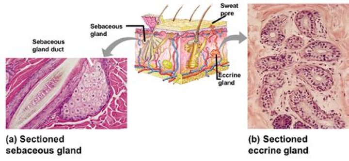
The pleura, which is a thin membrane that covers the inner surfaces of the thoracic cavity, consists of a layer of mesothelial cells supported by a network of connective and fibroelastic tissue. The visceral pleura lines the lung, whereas the parietal pleura lines the rib cage, diaphragm, and mediastinal structures.
Related Question Answers
What is pleura in human body?
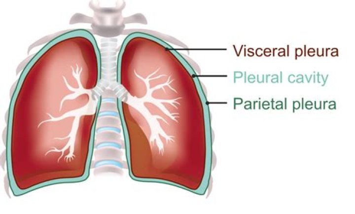
The pleura is a serous membrane which folds back onto itself to form a two-layered membrane structure. The thin space is known as the pleural cavity and contains a small amount of pleural fluid (few milliliters in a normal human). The outer pleura is attached to the chest wall (1-9).Where is pleura in the body?
Pleura, plural pleurae, or pleuras, membrane lining the thoracic cavity (parietal pleura) and covering the lungs (visceral pleura). The parietal pleura folds back on itself at the root of the lung to become the visceral pleura. In health the two pleurae are in contact.What is parietal pleura in anatomy?
The parietal pleura is the outer membrane which is attached to the inner surface of the thoracic cavity. It also separates the pleural cavity from the mediastinum. The parietal pleura is innervated by the intercostal nerves and the phrenic nerve. Between the membranes is a fluid-filled space called the pleural space.What cells are in the pleura?
The pleural mesothelial cell (PMC) is the most common cell in the pleural space and is the primary cell that initiates responses to noxious stimuli (3). PMCs are metabolically active cells that maintain a dynamic state of homeostasis in the pleural space.What are the lines of pleural reflection?
lines, usually projected onto the surface of the thoracic wall, indicating the abrupt change in direction of the parietal pleura as it passes from one wall of the pulmonary cavity to another. See also: vertebral line of pleural reflection.What is the mediastinal pleura?
Listen to pronunciation. (MEE-dee-uh-STY-nul PLOOR-uh) The thin membrane that lines the chest cavity in the area between the lungs.How is carbon dioxide removed from the lungs?
Blood rich in carbon dioxide then returns to the heart via the veins. From the heart, this blood is pumped to the lungs, where carbon dioxide passes into the alveoli to be exhaled.Does the pleural cavity contain the heart?
Thoracic cavity: The chest; contains the trachea, bronchi, lungs, esophagus, heart and great blood vessels, thymus gland, lymph nodes, and nerve,. Pleural cavities: Surround each lung. Pericardial cavity: Contains the heart. The pleural cavities flank the pericardial cavity.Does the pleural cavity contain the lungs?
The pleural cavity is surrounded by the rib cage, and itself surrounds the lungs. A small amount of fluid lies in the potential space between the two pleural layers.What is the pericardium?
The pericardium is a membrane, or sac, that surrounds your heart. It holds the heart in place and helps it work properly. Problems with the pericardium include. Pericarditis - an inflammation of the sac.Why is parietal pleura sensitive to pain?
The neurovascular supply differs for both layers of the pleura. The innervation of the parietal pleura is provided through the intercostal nerves (innervate the costal and cervical pleura), which causes it to be sensitive to pain, pressure and temperature.Is pleura a serous membrane?
Pleura. Pleurae are serous membranes that separate the lungs and the wall of the thoracic cavity. The visceral pleura covers the surface of the lungs, and the parietal pleura covers the inside of the thorax, mediastinum, and diaphragm. A thin film of serous fluid fills the space between the two pleurae.Where is pericardium found?
What is the pericardium? The pericardium is a thin sac that surrounds your heart. It protects and lubricates your heart and keeps it in place within your chest. Problems can occur when the pericardium becomes enflamed or fills with fluid.What is inside the pericardial cavity?
The pericardial cavity is the potential space formed between the two layers of serous pericardium around the heart. Normally, it contains a small amount of serous fluid that acts to reduce surface tension and lubricate. Therefore, the cavity facilitates the free movement of the heart.Why are the pleural membranes important for breathing?
Pleural Membrane FunctionThe pleural fluid also provides surface tension, keeping the lung suitably close to the wall of the thorax, despite the lungs not being directly fixed to it. The pleurae thus allow the volume of the lungs to change with the volume of the thoracic cavity, enabling ventilation.