Actin filaments are made up of identical actin proteins arranged in a long spiral chain. Like microtubules, actin filaments have plus and minus ends, with more ATP-powered growth occurring at a filament's plus end (Figure 2). Similarly one may ask, how are actin filaments formed?
(A) Actin monomers (G actin) polymerize to form actin filaments (F actin). The first step is the formation of dimers and trimers, which then grow by the addition of monomers to both ends. The actin monomers also bind ATP, which is hydrolyzed to ADP following filament assembly.
Secondly, what are intermediate filaments made up of? Whereas actin filaments and microtubules are polymers of single types of proteins (actin and tubulin, respectively), intermediate filaments are composed of a variety of proteins that are expressed in different types of cells.
Also to know is, what is actin filament and its function?
Several biological processes related to cell shape and movement depend on actin filaments (reviewed in [1]). In muscle cells, actin filaments are aligned and myosin proteins generate forces on the filaments to support muscle contraction.
What are actin filaments quizlet?
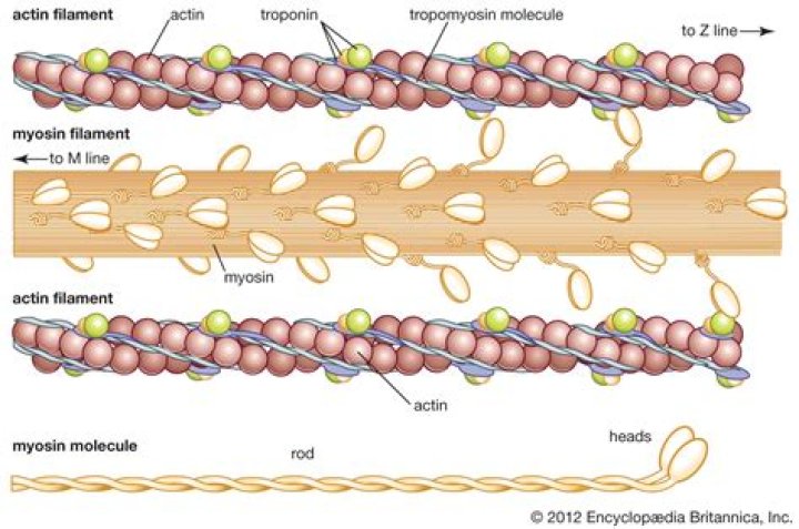
Actin filaments are polymers of actin monomers (G-actin). Actin filaments form the core of thin filaments in muscle cells. motor molecules that use ATP to pull on actin. Polymers of myosin in muscle cells are termed thick filaments.
Related Question Answers
Where are actin filaments found?
In many types of cells, networks of actin filaments are found beneath the cell cortex, which is the meshwork of membrane-associated proteins that supports and strengthens the plasma membrane. Such networks allow cells to hold — and move — specialized shapes, such as the brush border of microvilli. Where do actin filaments originate?
The evolutionary origin of actin can be traced to prokaryotic cells, which have equivalent proteins. Actin homologs from prokaryotes and archaea polymerize into different helical or linear filaments consisting of one or multiple strands. Is Actin a thick or thin filament?
Muscles are composed of two major protein filaments: a thick filament composed of the protein myosin and a thin filament composed of the protein actin. Muscle contraction occurs when these filaments slide over one another in a series of repetitive events. Where is actin and myosin produced?
Structure of muscle cells. Muscles are composed of bundles of single large cells (called muscle fibers) that form by cell fusion and contain multiple nuclei. Each muscle fiber contains many myofibrils, which are bundles of actin and myosin filaments organized (more) Who discovered actin?
Straub
Is there actin in the nucleus?
Actin, a globular protein with an approximately 42 kDa molecular weight, is found in all eukaryotic cells as one of the most highly-conserved proteins of the cytoskeleton. Recent evidence shows the association of actin with multiple nuclear complexes, thus the existence of actin in the nucleus is slowly being accepted. Are actin filaments dynamic?
Within cells, the actin cytoskeleton is dynamic, with filaments able to grow and shrink rapidly. The assembly, length, and stability of actin filaments are controlled by specialized actin-binding proteins. These proteins are in turn regulated by various mechanisms. What does actin mean?
Actin, protein that is an important contributor to the contractile property of muscle and other cells. It exists in two forms: G-actin (monomeric globular actin) and F-actin (polymeric fibrous actin), the form involved in muscle contraction. muscle: actin and myosin. What is the main function of actin?
These properties, along with its ability to transition between monomeric (G-actin) and filamentous (F-actin) states under the control of nucleotide hydrolysis, ions, and a large number of actin-binding proteins, make actin a critical player in many cellular functions, ranging from cell motility and the maintenance of Are Myofibrils?
A myofibril (also known as a muscle fibril) is a basic rod-like unit of a muscle cell. Muscles are composed of tubular cells called myocytes, known as muscle fibres in striated muscle, and these cells in turn contain many chains of myofibrils. What is the function of mitochondria?
Mitochondria are membrane-bound cell organelles (mitochondrion, singular) that generate most of the chemical energy needed to power the cell's biochemical reactions. Chemical energy produced by the mitochondria is stored in a small molecule called adenosine triphosphate (ATP). What is a Myofibril?
A myofibril is a long cylindrical organelle found in muscle cells formed by two transverse filament systems: the thick and thin filaments. The thin filament is composed primarily of actin; it is tethered at one end to the Z-disk, and it interdigitates with the thick filaments. What is the structure of actin?
The actin structure is highly conserved. The actin monomer consists of two major domains each of which contains two subdomains. The four subdomains are organized to form a rather flat molecule. Two large clefts are formed between the two major domains of actin. What is a sarcomere?
A sarcomere is the basic contractile unit of muscle fiber. Each sarcomere is composed of two main protein filaments—actin and myosin—which are the active structures responsible for muscular contraction. The most popular model that describes muscular contraction is called the sliding filament theory. What type of protein is tubulin?
Tubulin in molecular biology can refer either to the tubulin protein superfamily of globular proteins, or one of the member proteins of that superfamily. α- and β-tubulins polymerize into microtubules, a major component of the eukaryotic cytoskeleton. What causes actin polymerization?
Actin polymerization is controlled by intracellular signals that are mediated by small GTPases of the Rho family. The switch between the GTP to the GDP state can change the activity of actin-binding proteins and promote or retard polymerization of actin filament and growth of spines. What is myosin function?
Myosins are a large super-family of motor proteins that move along actin filaments, while hydrolyzing ATP to forms of mechanical energy that can be used for a variety of functions such as muscle movement and contraction. What is the main function of intermediate filaments?
Perhaps the most important function of intermediate filaments is to provide mechanical support for the plasma membrane where it comes into contact with other cells or with the extracellular matrix. Unlike microfilaments and microtubules, intermediate filaments do not participate in cell motility. What are examples of intermediate filaments?
The intermediate filaments are diverse; some 65 separate genes in humans have been identified. They all consist of three parts: a “head,” a long rod-like central part, and a “tail.” Examples of intermediate filaments include vimentin, desmin, glial fribrillary acid protein (GFAP), neurofilaments, and nuclear laminins. Are intermediate filaments made of keratin?
Keratin proteins comprise the two largest classes of intermediate filament proteins. The keratin filaments anchor the skin cells to the extracellular matrix (ECM) at their base and to adjacent cells at their sides, through structures called hemidesmosomes and desmosomes, respectively. How do intermediate filaments grow?
First, two monomers associate via their central domains to form parallel helical coils around each other. Intermediate filaments are built from monomers that associate with each other form dimers. Pairs of dimers then associate in an anti-parallel fashion to form staggered tetramers. What is cellular motility?
Definition. Cellular motility is the spontaneous movement of a cell from one location to another by consumption of energy. The term encompasses several types of motion, including swimming, crawling, gliding and swarming. Do sperm cells have intermediate filaments?
Evidence from indirect immunofluorescence microscopy, immunoblotting, and partial peptide analysis indicates that the intermediate filament proteins vimentin and keratin are present in human sperm cell acrosomes. What type of filament is collagen?
The first intermediate filament (IF) proteins studied were keratins. The name keratin is derived from the Greek word for horn: κερας. They are—like the non-IF proteins myosin, fibrinogen, and collagen—abundant and highly insoluble constituents of metazoan cells and tissues. Are Cytoskeletons in plant cells?
Abstract. The eukaryotic cytoskeleton is a dynamic filamentous network with various cellular and developmental functions. Plant cells display a singular architecture, necessitating a structurally and functionally unique cytoskeleton and plant specific control mechanisms. What cells have a cytoskeleton?
Cytoskeleton, a system of filaments or fibres that is present in the cytoplasm of eukaryotic cells (cells containing a nucleus). Are actin filaments and Microfilaments the same thing?
Of the three types of protein fibers in the cytoskeleton, microfilaments are the narrowest. They function in cellular movement, have a diameter of about 7 nm, and are made of two intertwined strands of a globular protein called actin (Figure 4.5. 2). For this reason, microfilaments are also known as actin filaments. Which of the following is the largest of the cytoskeletal elements?
Microtubules
Which is the main function of intermediate filaments quizlet?
What is the main function of intermediate filaments? Intermediate filaments have great tensile strength, and their main function is to enable cells to withstand the mechanical stress that occurs when cells are stretched. What is the role of actin in eukaryotic cells quizlet?
What is the role of actin in eukaryotic cells? It provides strength in stretching and compression of a cell. responsible for cellular respiration. Is the motor protein involved in the movement of eukaryotic flagella?
In flagella and motile cilia, motor proteins called dyneins move along the microtubules, generating a force that causes the flagellum or cilium to beat.